Muscles Labeled Front And Back / GCSE PE - Muscular System | Teaching Resources - C rnrceps brachn l unssimus dorsi k.
Dapatkan link
Facebook
X
Pinterest
Email
Aplikasi Lainnya
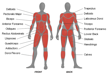
Muscles Labeled Front And Back / GCSE PE - Muscular System | Teaching Resources - C rnrceps brachn l unssimus dorsi k.. Want to learn more about it? Muscles vary greatly in their shape and size. Label muscles front and back view. The external intercostal muscles, or external intercostals (intercostales externi) are eleven in number on both sides. Label the following anatomicalsites in the diagram:
Back view of muscles, skeleton, organs, nervous system. 12 photos of the muscles labeled front and back. The muscles of the back that work together to support the spine, help keep the body upright and allow twist and bend in many directions. This labeled human muscular system chart illustrates the major muscle groups in the back (posterior) view and the front (anterior) view. What do you prefer to learn with?
Triathlons and beyond.: Physiotherapy Day 1. from 2.bp.blogspot.com Each of your muscles is made up of thousands of thin, long, cylindrical cells called muscle fibers. These muscles are able to move the upper limb as they originate at the vertebral column and insert onto. What do you prefer to learn with? Muscle imbalances not only look odd but can increase your risk of injury. Triceps, biceps, pectoralis major, quadriceps , hamstrings, gluteus maximus , abdominals, deltoid, latissimus dorsi, external obliques, gastrocnemius , tibialis anterior. Barbell back squat the barbell back squat focuses more on the hips, glutes and lumbar spine and places more of the load on the posterior half of your body. More specifically, this beautifully illustrated anatomy chart includes neck and shoulders, multiple views of the back and spine, and frontal views of each muscular extremity of the human body. The muscle fibers' highly specialized structure enables the muscles to relax and contract to produce movement.
This muscular system chart shows in detail the deep layers of muscle on the back side of your body.
What is the primary action of the muscle labeled 6? What do you prefer to learn with? The trapezius originates from the skull and spine of the. Proportionate development of the upper and lower and front and back parts of your body. The muscles extend from the tubercles of the ribs behind, to the cartilages of the ribs in front, where they end in thin membranes, the external intercostal membranes. C rnrceps brachn l unssimus dorsi k. Label muscles front and back view. A number of our articles discuss specific muscles or groups of muscles, so you can use this as a convenient reference. Rotator cuff muscle with anatomical posterior and anterior view expample. This muscular system chart shows in detail the deep layers of muscle on the back side of your body. A back muscle that pulls the arm down and back. Within this group of back muscles you will find the latissimus dorsi, the trapezius, levator scapulae and the rhomboids. The superficial back muscles are the muscles found just under the skin.
Label muscles front and back view. Muscle imbalances not only look odd but can increase your risk of injury. The superficial back muscles are the muscles found just under the skin. What is the primary action of the muscle labeled 6? Want to learn more about it?
11 best Muscles/Labeled images on Pinterest | Physical ... from i.pinimg.com Learn how to identify, fix, and prevent them in this article. Back view of muscles, skeleton, organs, nervous system. The trapezius originates from the skull and spine of the. Label the following anatomicalsites in the diagram: Proportionate development of the upper and lower and front and back parts of your body. The main muscles involved are your glutes, quads, hamstrings, lower back and calves and you'll activate your core for stabilizing purposes as well. A back muscle that pulls the arm down and back. Tutorials and quizzes on the anatomy and actions of the back muscles (iliocostalis, longissimus, spinalis, multifidus, and quadratus lumborum), using interactive animations, diagrams, and illustrations.
Label the following anatomicalsites in the diagram:
The trapezius originates from the skull and spine of the. The biggest muscle is lats muscle, then there are traps muscle. There are two parallel muscles. The muscle fibers' highly specialized structure enables the muscles to relax and contract to produce movement. Within this group of back muscles you will find the latissimus dorsi, the trapezius, levator scapulae and the rhomboids. Identify the muscle labeled 8. This muscular system chart shows in detail the deep layers of muscle on the back side of your body. Start studying muscle labeling (front view). The superficial back muscles are the muscles found just under the skin. The muscles of the back that work together to support the spine, help keep the body upright and allow twist and bend in many directions. Rotator cuff muscle with anatomical posterior and anterior view expample. Attachments, nerve supply well there are lot of muscles on back and every muscle is trained differently. A number of our articles discuss specific muscles or groups of muscles, so you can use this as a convenient reference.
Back of the head muscle structure and nerve system diagram. More specifically, this beautifully illustrated anatomy chart includes neck and shoulders, multiple views of the back and spine, and frontal views of each muscular extremity of the human body. A back muscle that pulls the arm down and back. This muscular system chart shows in detail the deep layers of muscle on the back side of your body. The superficial back muscles are the muscles found just under the skin.
Health and Fitness | Body by Wright Blog | Page 3 from bodybywright.files.wordpress.com These muscles are able to move the upper limb as they originate at the vertebral column and insert onto. Back of the head muscle structure and nerve system diagram. Barbell back squat the barbell back squat focuses more on the hips, glutes and lumbar spine and places more of the load on the posterior half of your body. What is the primary action of the muscle labeled 6? Back view of muscles, skeleton, organs, nervous system. Intermediate back muscles and c. Which muscles contract and which muscles relax when you turn your head to the right? The external intercostal muscles, or external intercostals (intercostales externi) are eleven in number on both sides.
Muscle imbalances not only look odd but can increase your risk of injury.
Start studying muscle labeling (front view). Learn vocabulary, terms and more with flashcards, games and other study tools. 12 photos of the muscles labeled front and back. The muscles extend from the tubercles of the ribs behind, to the cartilages of the ribs in front, where they end in thin membranes, the external intercostal membranes. This muscular system chart shows in detail the deep layers of muscle on the back side of your body. The muscle fibers' highly specialized structure enables the muscles to relax and contract to produce movement. A large, complex group of muscles work together to attached to the front of the spine, these muscles include the abdominal muscles. The external intercostal muscles, or external intercostals (intercostales externi) are eleven in number on both sides. Male muscular system, full anatomical body diagram with muscle scheme, vector illustration educational poster. Aalso known as the six pack, is a paired muscle running vertically on each side of the front wall of the abdomen. Intermediate back muscles and c. The anterior muscles of the torso (trunk) are those on the front of the body, including the muscles of the chest, abdomen, and pelvis. Muscle imbalances not only look odd but can increase your risk of injury.


Komentar
Posting Komentar